【印刷可能】 sinus tachycardia with pvcs. no st segment elevation 117587-Do pvcs cause tachycardia
Sinus tachycardia, rSR' in V 1 –V 2, right ventricular hypertrophy, rightward QRS axis shift,"P pulmonale" Rapid regression of abnormalities on serial tracings favors pulmonary embolism rather than myocardial infarction Hypokalemia STsegment depression Twave flattening Prominent U wave (with the flattened T wave, may mimic a wide and notched upright T wave) Prolonged QTU · Sinus tachycardia is a regular cardiac rhythm in which the heart beats faster than normal and results in an increase in cardiac output While it is common to have sinus tachycardia as a compensatory response to exercise or stress, it becomes concerning when it occurs at rest1 The normal resting heart rate for adults is between 60 and 100, which varies based on the level/03/18 · The ECG showed widecomplex tachycardia with a rate in the low 0s as shown in Figure 1A Given the patient's stable blood pressure, he was started on procainamide therapy with an initial 100 mg intravenous bolus followed by an infusion Within minutes, the patient converted to sinus rhythm with resolution of his symptoms Repeat ECG, however, was now concerning for lateral STsegment
St Segment Elevation In Lead Avr Is This A Stemi Equivalent Ems 12 Lead
Do pvcs cause tachycardia
Do pvcs cause tachycardia-ST segment elevation is present Accelerated idioventricular rhythm Normal sinus rhythm with firstdegree AV block and one nonconducted PAC (follows the fifth QRS complex) Sinus tachycardia Sinus bradycardia;The ST elevation in V26 is concave upwards, another
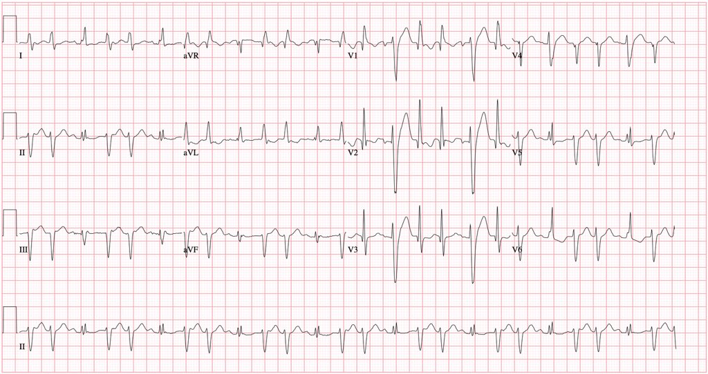


Jason Andrade This Ecg Demonstrates Complete Heart Block With A Very Slow Escape Rhythm There Is A Clear Escape In The Middle Of The Twcibg But Right At The Beginning
This encounter shows a fast rate over 100 bpm, with a regular rhythm and P waves, indicating sinus tachycardia The extremely long delay between the P wave and QRS indicates first degree heart block Additionally, ectopy is seen in this encounter, in the form of premature ventricular contractions These wide, bizarre looking QRS complexes canHowever, myocardial necrosis occurs once again from coronary artery occlusion · This ECG shows an underlying rhythm of normal sinus rhythm at a rate of 80 / min There are two premature ventricular contractions (PVCs) The sinus rhythm actually continues uninterrupted, causing a "compensatory pause" If you march out the P waves, you may even see hints of the hidden P waves in the ST segments of the PVCs The P waves that occur in the ST segments of the PVCs
· ST segment elevation Transient ST segment elevation in patients with chest pain is a feature of ischaemia and is usually seen in vasospastic (variant or Prinzmetal's) angina A proportion of these patients, however, will have substantial proximal coronary artery stenosis When ST segment elevation has occurred and resolved it may be followed by deep T wave inversion · Subcutaneous crepitus was not observed An ECG performed on the same day (Fig 2A) demonstrated sinus tachycardia with up to 3 mm (03 mV) STsegment elevation in the inferior leads and a somewhat unusual STsegment elevation in precordial leads V 3 –V 5 An ECG 4 h later (Fig 2B) continued to show STsegment elevation The interpretation80% vs 41%, p = 001) The combination of these ECG findings identified TC with a specificity of 95% and a positive predictive value of
Sinus bradycardia with one interpolated PVC; · 1 Am J Cardiol 01 Aug 15;(4)4147 Usefulness of sinus tachycardia and STsegment elevation in V(5) to identify impending left ventricular free wall rupture in inferior wall myocardial infarctionSupraventricular tachycardia may also cause ST segment depressions These depressions are usually horizontal or upsloping and tend to be most evident in leads V4–V6 these ST segment depressions resolve rapidly once the tachycardia has resolved A novel cardiac syndrome with concaveupward STsegment depressions
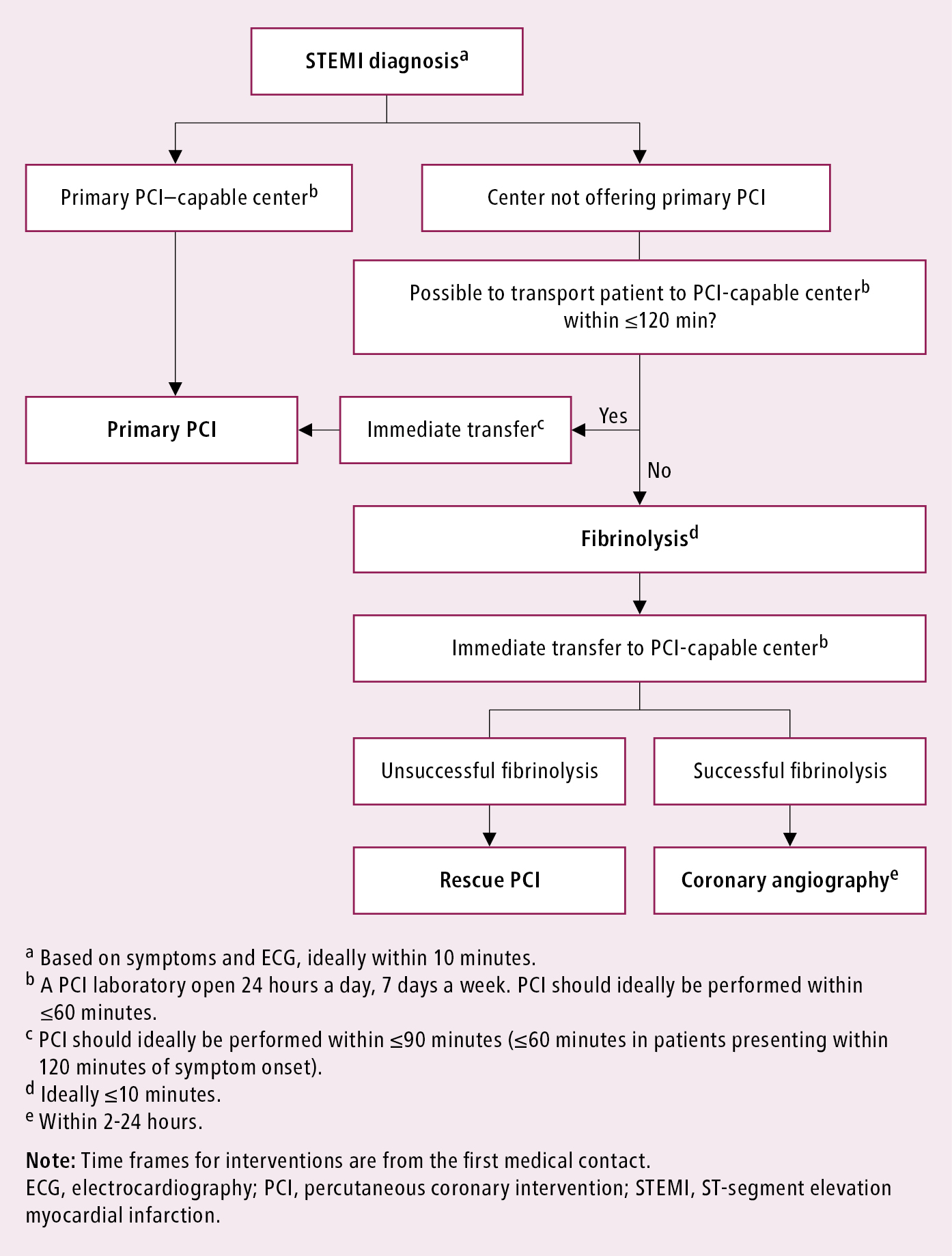


St Segment Elevation Myocardial Infarction Stemi Acute Coronary Syndrome Acs Ischemic Heart Disease Ihd Cardiovascular Diseases Diseases Mcmaster Textbook Of Internal Medicine
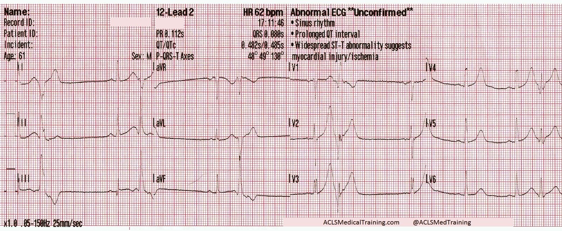


Chest Pain And Transient St Segment Elevation Acls Medical Training
· ECG activity was recorded and classified as follows normal sinus rhythm, sinus tachycardia, sinus bradycardia, atrial fibrillation (AFib), atrial flutter, STsegment depression, STsegment elevation, multifocal atrial tachycardia (MAT), premature atrial contraction (PAC), premature ventricular contraction (PVC), atrioventricular block (AVB), left bundle branch blockSinus Tachycardia ECG (Example 1) Anterior Wall ST Segment Elevation Myocardial Infarction (MI) ECG (Example 1) Anterior Wall ST elevation MI ECG (Example 2) Anterior Wall ST elevationVentricular tachycardia is a fast heart rate, anything over the normal 100 beats per minute, which starts in the lower chambers of the heart, the ventricles It causes the ventricles to contract before they have had a chance to completely fill with blood, impairing blood flow to the body Premature Ventricular Contractions (PVCs) are single


The Six Second Ecg Squarespace Pages 1 6 Flip Pdf Download Fliphtml5


Ecg Shows Sinus Tachycardia With Widespread St Elevation Pr Elevation Download Scientific Diagram
· This is ST elevation of myocardial infarction A follow up ECG about 15 minutes later was nearly identical except that there was no PVC I have estimated the QTc as 418ms If you do not recognize this as STEMI, you can use the formula (STE60V3 = 25, QT = 418, RAV4 = 55) and you get 2585 which is pretty much diagnostic of STEMIDifferential Diagnosis of ST Segment Elevation Normal Variant "Early Repolarization" (usually concave upwards, ending with symmetrical, large, upright T waves) Example #1 "Early Repolarization" note high take off of the ST segment in leads V46;I went into to a preop appointment had a heart rate of 147 they did ekg that says sinus tachycardia and borderline st elevation, anterior leads hr147 pr1 qrsd 72 qt251 qtc393 they made it seem like a big worry but didnt tell me what to do?
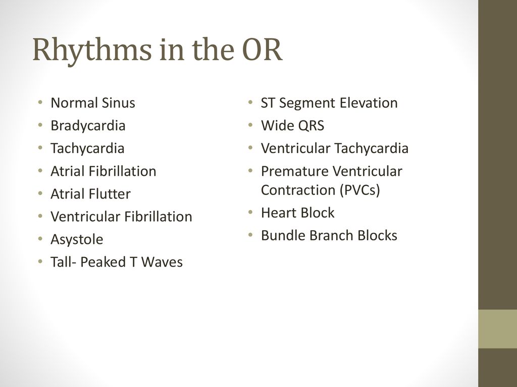


Ecgs For Perfusion Michael F Hancock Ccp Cooper University Hospital Ppt Download


Treatment Of Ventricular Arrhythmias The Cardiology Advisor
· Sinus bradycardia is the opposite of sinus tachycardia and happens when your sinus node doesn't send enough impulses, resulting in a heart rate of fewer than 60 beats per minuteThe ST elevation in V23 is generally seen in most normal ECG's;Sinus tachycardia is recognized on an ECG with a normal upright P wave in lead II preceding every QRS complex, indicating that the pacemaker is coming from the sinus


Sinus Tachycardia And Diffuse St Segment Elevation Suggestive Of Early Download Scientific Diagram


Pdf Heart Rhythm Disorders And Myocardial Remodeling In Patients With St Segment Elevation Acute Myocardial Infarction Semantic Scholar
The T wave during sinus tachycardia or if the PR segment is depressed or there is a prominent atrial repolarization (Ta) wave Tracing 1 in Figure 1 is an example of normal STsegment elevation In a study of 6014 healthy men in the US Air Force who were 16 to 58 years old, 91 percent had STsegment elevation of 1 to 3 mm in one or more precordial leads 6 The elevation was most/03/21 · It is accompanied by STsegment and T wave changes every second sinus beat is followed by a PVC Ventricular quadrigeminy every third sinus beat is followed by a PVC Couplet two consecutive PVCs Nonsustained ventricular tachycardia three or more consecutive PVCs Ventricular bigeminy premature ventricular complexes alternating with a normal beat Three or more consecutive PVCsBelow are Examples of ST Segment Elevation More on PVCs 3 or more PVC's in a row can be called Ventricular Tachycardia Interpretation NSR into Paroxysmal Venticular Tachycardia Ventricular Rhythms Idioventricular Rhythm is referred to as IVR NOTE In the interpretation always state the underlying rhythm For example, the rhythm above would be interpreted this


Electrocardiogram Ecg Shows Sinus Tachycardia St Segment Elevation Download Scientific Diagram


Ecg Learning Center An Introduction To Clinical Electrocardiography
· Sinus tachycardia refers to a fasterthanusual heart rhythm Your heart has a natural pacemaker called the sinus node, which generates electrical impulses that move through your heart muscle and · Sinus tachycardia refers to an increased heart rate that exceeds 100 beats per minute (bpm) The sinus node, or sinoatrial node, is a bundle of · More often, tachycardia with ST segment abnormalities (elevation or depression) is due to an underlying illness (PE, sepsis, hemorrhage, dehydration, hypoxia, respiratory failure, etc) One must clearly rule out these processes before jumping on the ACS diagnosis
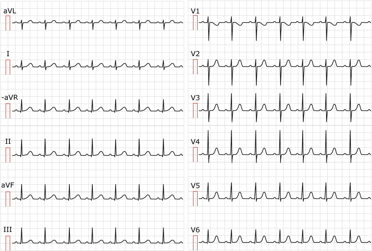


Sinus Tachycardia Inappropriate Sinus Tachycardia Ecg Echo


Cmc Compendium Carolinas Ekg Blog
· 60 Mugnai et al "The absence of abnormal Q waves, the ST depression in aVR and the lack of ST elevation in V1 were significantly associated with TC (respectively 52% vs 18%, p = 001; · ECG Findings The rhythm is sinus tachycardia, at a rate of 1 bpm The QRS is narrow at 08 seconds ( ms) While the PR interval is normal, at 14 seconds (140 ms), the PR segment is very short The PR segment is the line between the end of the P wave to the beginning of the QRS complex This can indicate the presence of an accessory pathway that bypasses the · There is ST segment elevation in all precordial leads, except for possibly V6 Every other sinus beat is obscured by the PVCs By the end of the strip, the underlying sinus rhythm has slowed slightly The ECG signs that the ectopic beats are ventricular are lack of P waves associated with the premature beats, QRS width about 16 seconds, and compensatory pauses The axis of the sinus
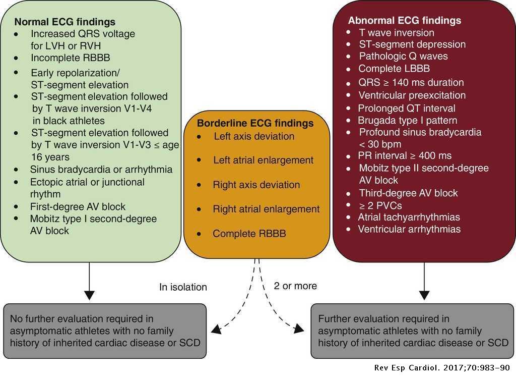


Comments On The New International Criteria For Electrocardiographic Interpretation In Athletes Revista Espanola De Cardiologia


St Segment Elevation In Lead Avr Is This A Stemi Equivalent Ems 12 Lead
47% vs 11%, p = 001;LongStanding sinus tachycardia may lead to STsegment depressions on the ECG Such STsegment depressions may be seen anywhere but most commonly in leads V3, V4, V5 and V6 The STsegment tends to be either horizontal or upsloping Longstanding sinus tachycardia may also cause diminished Twave amplitude on the ECGDr Murray Wellner answered 45 years experience Internal Medicine Rule out CAD The rate is much to fastSinus tachycardia


Premature Ventricular Contractions Pvcs Induced By Administration Of Cilostazol After Myocardial Infarction Russian Open Medical Journal


Basic Electrocardiography Guide To Diagnostic Tests
V2–V3 ≥0,02 s or QS complex* None All other leads ≥0,03 s and ≥1 mm deep (or QS complexThe above is a normal sinus rhythm with a rate of 68 beats per minute with ST elevation of 2 mm (2 small boxes) The arrow is pointing at the ST segment of the EKG ST elevation in certain leads can be normal, so a thorough evaluation would require a 12 lead EKG and perhaps additional tests if the patient is symptomatic Symptoms may include chest tightness, pressure or pain and/or · The ECG shows sinus tachycardia with diffuse STsegment elevation in all leads and STsegment depression in lead aVR Echocardiogram showed severe left ventricular dysfunction with apical hypokinesia Coronary angiogram showed mild plaquing in the proximal left anterior descending artery with the rest being normal Spontaneous coronary artery dissection should be
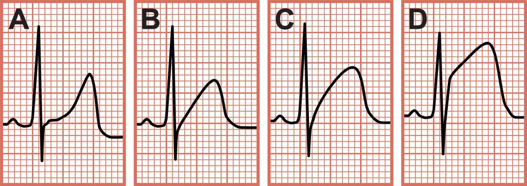


Inferior Wall Myocardial Infarction Chapter 2 Critical Cases In Electrocardiography


Introduction To Telemetry Monitoring For Arrhythmias Nursing Ce Course Nursingce
The ICD10CM code R9431 might also be used to specify conditions or terms like ambulatory ecg abnormal, anterior and lateral st segment elevation, anterior myocardial infarction on electrocardiogram, anterior st segment depression, anterior st segment elevation ,ST segment elevation in leads V1–V2 All degrees of AV block Sinus node dysfunction and tachbrady syndrome Ventricular tachycardia, ventricular fibrillation and torsade de pointes Hypocalcaemia Etiology of hypocalcaemia Acute pancreatitis, pancreas surgery, alkalosis (hyperventilation), rhabdomyolysis, septicemia (sepsis), osteolytic cancer metastases, abnormalIn patients with STEMI, STsegment elevations and pathological Qwaves occur in the same leads, which is why pathological Qwaves can be used to localize the infarct area ECG criteria for pathological Qwaves (Qwave infarction) Lead Definition of pathological Qwave Normal variants;


Premature Ventricular Contraction Wikipedia


Ekg Nov 16 Nov
A nonST segment elevation myocardial infarction occurs when there is no ST segment elevation on the ECG;Sinus bradycardia occurs on an ECG when there is a normal upright P wave in lead II ― sinus P wave ― preceding every QRS complex with a ventricular rate of less than 60 beats per minute · ST‐segment elevation during arrhythmia ablations—A review supraventricular tachycardia ablations, coronary sinus ablation, and ventricular arrhythmia ablations In this review, we have performed a literature search and attempt to provide the readership with a comprehensive overview of ST segment elevation during ablations and its mechanism and guidance for
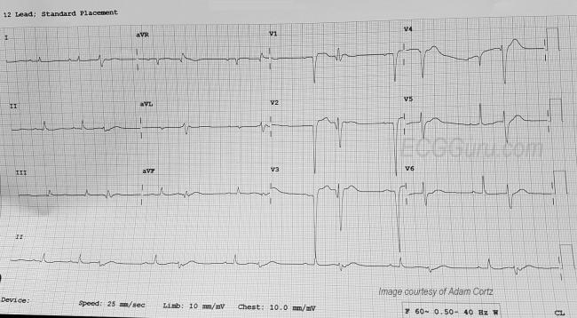


Anterior Wall M I With Ventricular Bigeminy Ecg Guru Instructor Resources
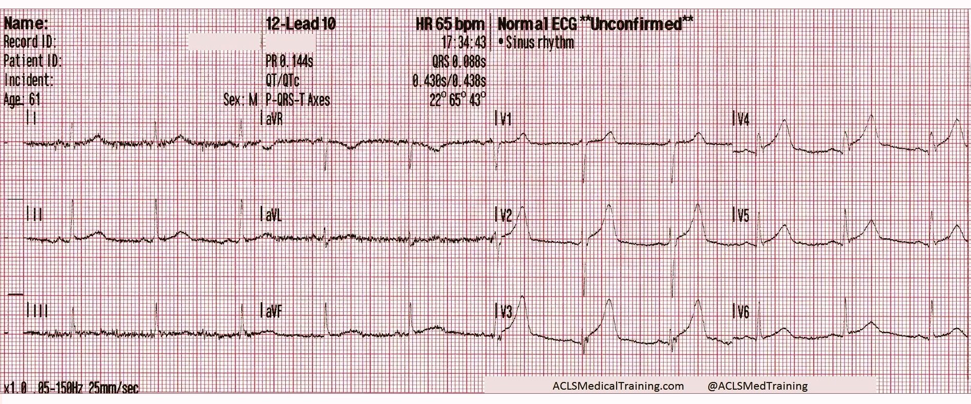


Chest Pain And Transient St Segment Elevation Acls Medical Training
· ECG No 1 @1743 The rhythm is sinus tachycardia at 118 bpm The There is ST segment elevation in all precordial leads, except for possibly V6 The shape of the ST segments in the anterior wall range from coved upward in a "frowning" shape (V1) to very straight (V5 and V6) There is also ST elevation in aVL with ST straightening in Lead I There is ST depression in theCardiac monitor in lead II demonstrated sinus bradycardia with ST segment elevation The patient then lost consciousness, and the cardiac monitor revealed ventricular fibrillation ( Figure 1A ) Immediate defibrillation at 0 joules restored a pulse with a rate of approximately 140 bpm Blood pressure was measured at 170/90 mm Hg · ST segment elevation during various ablation procedures is frequently being recognized The mechanism of STE is not exactly known, however, there are various possible mechanisms postulated for these ECG changes As was previously eluded, some of these mechanisms include direct mechanical injury, coronary spasm, thermal injury, ganglionic plexus
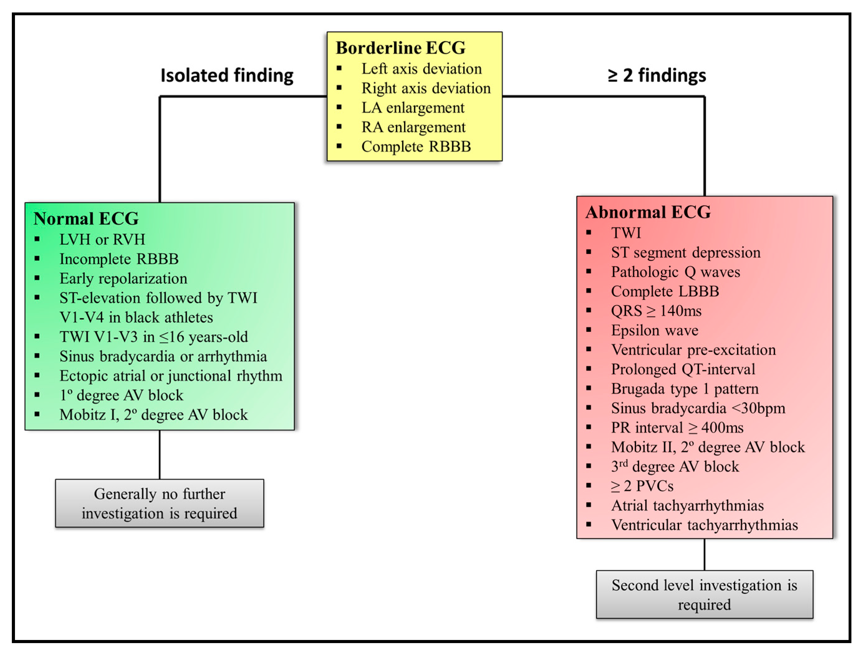


Medicina Free Full Text Sudden Cardiac Death In Athletes From The Basics To The Practical Work Up Html


The Electrocardiography Shows A Sinus Tachycardia And St Segment Download Scientific Diagram
/07/ · Cases by Type Select Type 21 AV Block 15 ECG Competition 15 ECG Competition Part II 16 ECG Competition 17 ECG Competition Part II 18 ECG Competition Part II 19 ECG Competition ECG Competition 5 Step Approach 5FU aberrancy Accelerated idioventricular rhythm Acidosis ACS ACS mimics ACS RIsk Factors Acute Pericarditis AdvancedIii) Atrial Flutter with 21 AV conduction;STsegment depression is present Accelerated junctional rhythm Atrial flutter with 41 AV conduction Normal sinus rhythm;


Ecg Shows Sinus Tachycardia With Widespread St Elevation Pr Elevation Download Scientific Diagram


Dr Smith S Ecg Blog Tachycardia And St Elevation
· The short horizontal YELLOW lines indicate the ST segment baseline There is no ST segment elevation for the PVC in lead V1 — which is what one normally expects in PVCs when there is no ongoing OMI However, it should be obvious that the PVCs in leads V2 and V3 manifest significant Jpoint elevation that justshouldn'tbethere · For practical purposes, when the ventricular response is this fast, the differential diagnosis consists of 4 entities i) Sinus tachycardia (which could still be present, if sinus P waves were hidden within the preceding STT wave);Patient has a possible inferior myocardial infarction Rhythm analysis indicates sinus tachycardia with ST segment elevation This encounter shows ST segment elevation, an often significant EKG finding ST elevation can be identified by looking at the J point, which should be elevated at least 01mV above the isoelectric line


Premature Ventricular Contraction An Overview Sciencedirect Topics
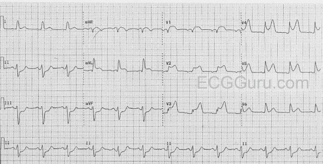


St Elevation Ecg Guru Instructor Resources
Ii) Reentry SVT (such as AVNRT or AVRT);We describe a 41yearold man who presented with steep downsloping STsegment elevation, paroxysmal ventricular tachycardia, severe sinus bradycardia, and intermittent complete ncbinlmnihgov This may be caused by a slowed signal from the sinus node ( sinus bradycardia ), by a pause in the normal activity of the sinus node (sinus arrest), or by blocking of theSinus bradycardia, two premature ventricular contractions, incomplete right bundle branch block and STsegment depressions in V2V6 Click to zoom Figure 3 Sinus bradycardia The small qwaves in inferior leads (II, aVF, III) are not significant Low voltage in limb leads Click to zoom Figure 4 Sinus bradycardia Click to zoom Treatment of sinus bradycardia general aspects of


The Six Second Ecg Squarespace Pages 1 6 Flip Pdf Download Fliphtml5


Ventricular Bigeminy Ecg Example 1 Learntheheart Com
And, iv) Atrial Tachycardia with 21 AV conduction Awareness of
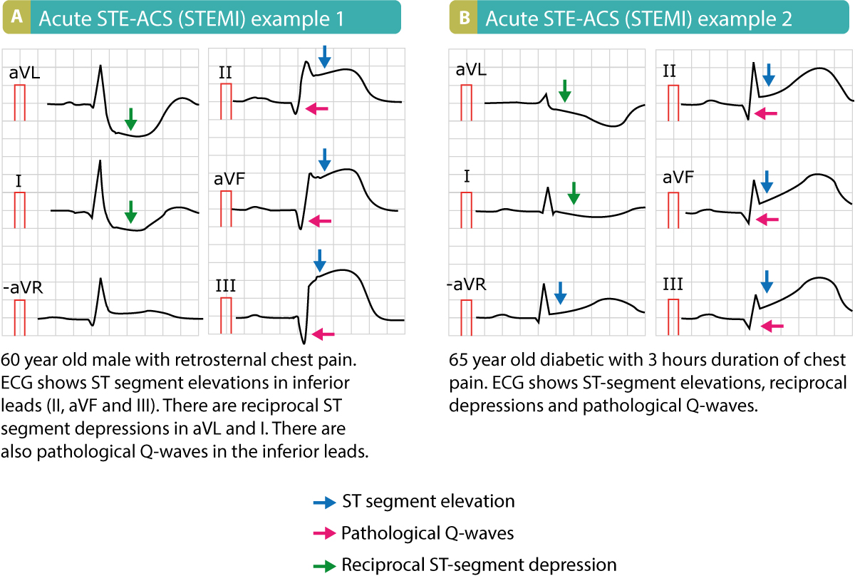


Stemi St Elevation Myocardial Infarction Diagnosis Criteria Ecg Management Ecg Echo


Dr Smith S Ecg Blog Tachycardia Dehydration And New St Elevation In A Something Then A Surprise


Dr Smith S Ecg Blog Chest Pain Sinus Tachycardia And St Elevation


Ecg Interpretation Of Arrhythmias Tusom Pharmwiki


Ecg Learning Center An Introduction To Clinical Electrocardiography
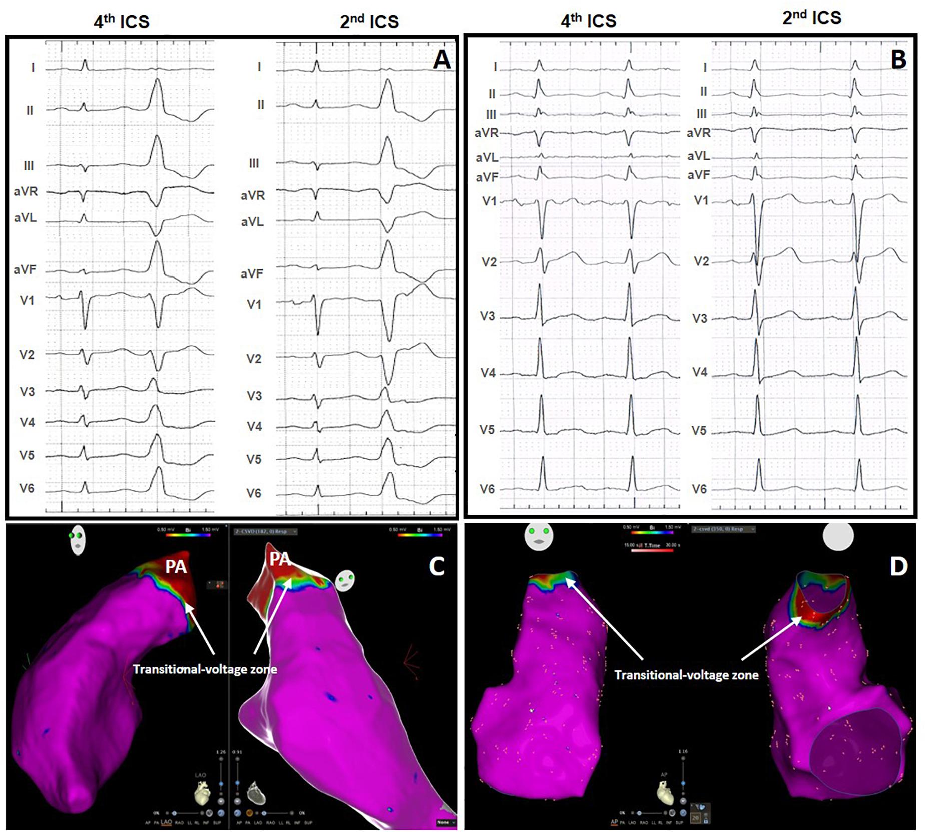


Frontiers Idiopathic Premature Ventricular Contractions From The Outflow Tract Display An Underlying Substrate That Can Be Unmasked By A Type 2 Brugada Electrocardiographic Pattern At High Right Precordial Leads Physiology


Ecg Quiz Accelerated Idioventricular Rhythm Arrhythmogenic Right Ventricular Dysplasia Arvd Ashman Phenomenon Asystole Atrial Fibrillation Atrial Flutter Atrial Tachycardia Atrioventricular Block Atrioventricular Dissociation Atrioventricular Nodal


Rare Case Of Selenite Poisoning Manifesting As Non St Segment Elevation Myocardial Infarction Jacc Case Reports


Cmc Ecg Monthly Session Emergency Medicine Guidewire


Premature Ventricular Contractions Pvcs Induced By Administration Of Cilostazol After Myocardial Infarction Russian Open Medical Journal


Non St Segment Elevation Myocardial Infarction Associated With Inadvertent Thyroid Hormone Overdose Abstract Europe Pmc


Dr Smith S Ecg Blog Chest Pain Sinus Tachycardia And St Elevation


St Changes Heart Squad


1


Jason Andrade This Ecg Shows Svt Degenerating Into Self Limited Polymorphic Vt Following Adenosine One Proposed Interpretation Was Svt Degenerating Into Af With Preexcitation But The Svt Is Likely Avnrt


Premature Ventricular Contraction Article


Electrophysiology Of The Heart Ecg Monitoring The Ecg
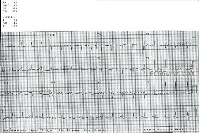


Sinus Tachycardia Ecg Guru Instructor Resources


Electrocardiography Wikidoc
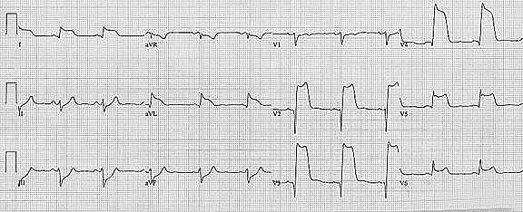


Tombstone St Elevation Wikidoc


1


Http Cardiolatina Com Wp Content Uploads 19 01 Study Methodology For Screening Candidates To Athletes Risk Pdf


Premature Ventricular Contractions Reassure Or Refer Cleveland Clinic Journal Of Medicine


Acls Rhythm Test 2 A 74 Year Old Woman With Chest Pain Blood Pressure 192 90 And Rates Her Pain 9 10 Pdf Free Download
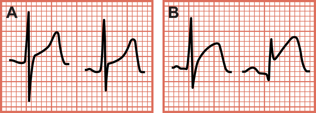


Confusing Conditions St Segment Elevations And Tall T Waves Coronary Mimics Chapter 7 Critical Cases In Electrocardiography


Electrocardiography Wikipedia


Jason Andrade This Ecg Demonstrates Complete Heart Block With A Very Slow Escape Rhythm There Is A Clear Escape In The Middle Of The Twcibg But Right At The Beginning


Acls Rhythm Test 2 A 74 Year Old Woman With Chest Pain Blood Pressure 192 90 And Rates Her Pain 9 10 Pdf Free Download
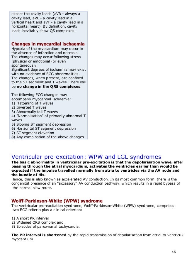


The Normal Ecg And The 12


St Segment Elevation During Arrhythmia Ablations A Review Kichloo 21 Journal Of Arrhythmia Wiley Online Library


Premature Ventricular Complex Pvc Litfl Ecg Library Diagnosis
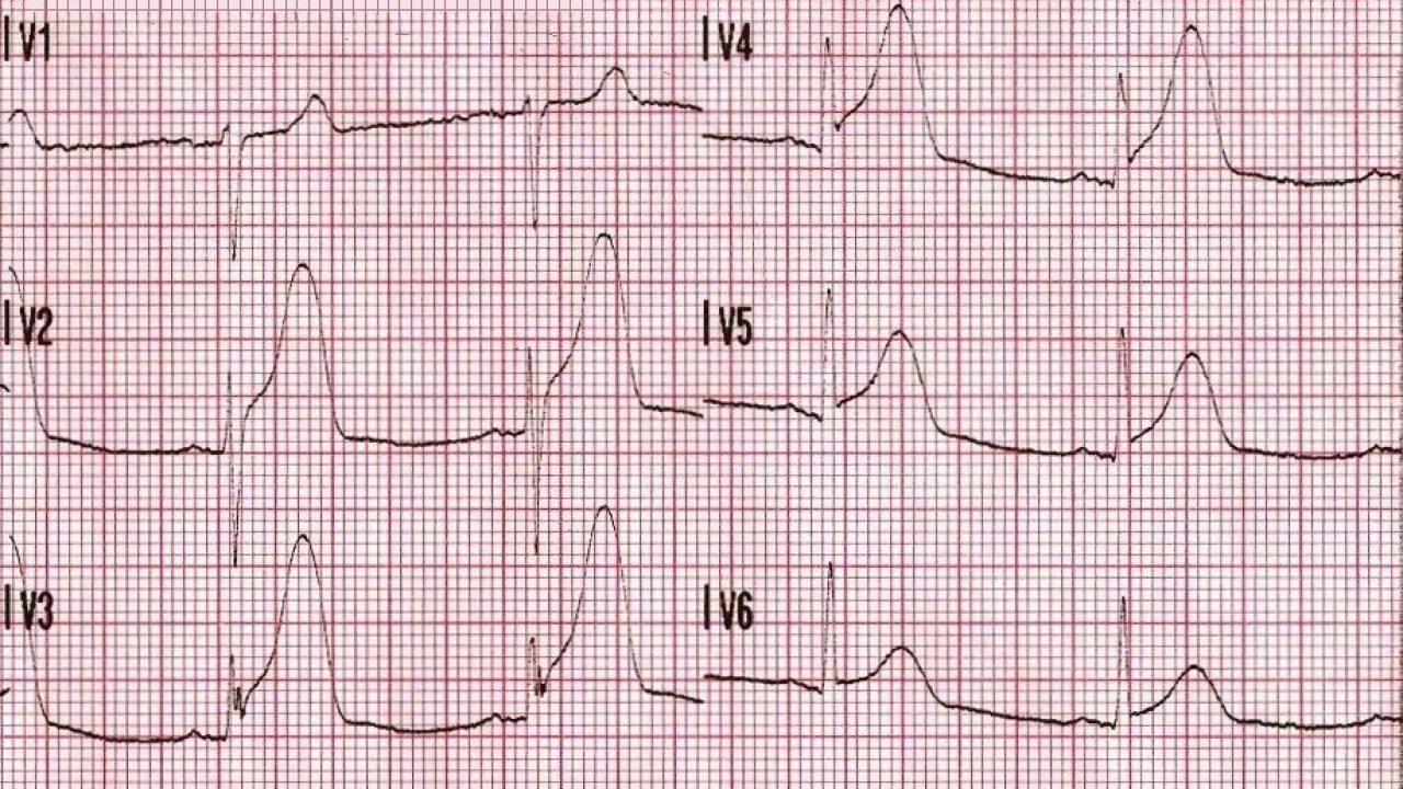


Chest Pain And Transient St Segment Elevation Acls Medical Training


St Segment Elevation And Depressions In Supraventricular Tachycardia Without Coronary Artery Disease


International Recommendations For Electrocardiographic Interpretation In Athletes Sciencedirect


1
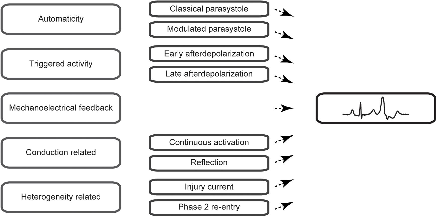


Frontiers Pathophysiological Mechanisms Of Premature Ventricular Complexes Physiology


Heart Rhythm Disorders And Myocardial Remodeling In Patients With St Segment Elevation Acute Myocardial Infarction
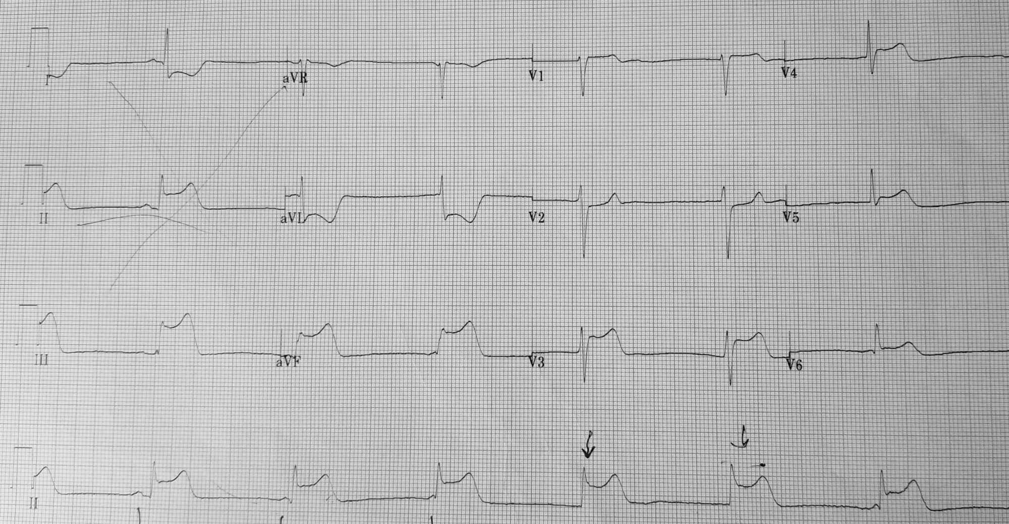


Cureus A Rare Case Of Vasospastic Angina Presenting With Inferior Lead St Segment Elevation And Ventricular Fibrillation In The Absence Of Coronary Obstruction A Case Report


Ecg Chp 6 Ventricular Rhythms Flashcards Quizlet


Www Ahajournals Org Doi Pdf 10 1161 Jaha 1 Download True


1
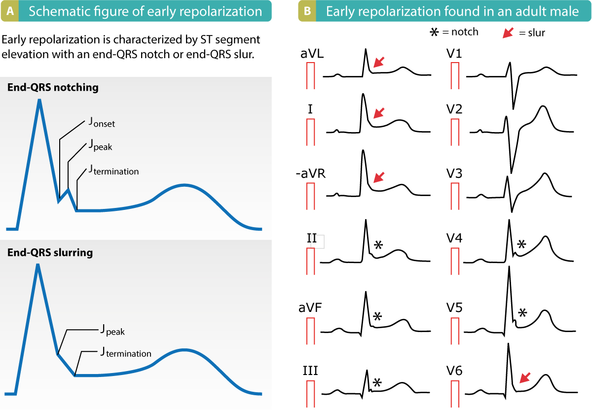


Early Repolarization Pattern On Ecg Early Repolarization Syndrome Ecg Echo


Dr Smith S Ecg Blog Chest Pain Sinus Tachycardia And St Elevation


Ekg Medic Rhythms Atrial And Vent Flashcards Quizlet


Premature Ventricular Contractions Reassure Or Refer Cleveland Clinic Journal Of Medicine


Premature Ventricular Contractions Pvcs Ecg Review Criteria And Examples Learntheheart Com


Sinoatrial Exit Block Sa Block N 2 Nd


Ekg Nov 16 Nov


Dr Smith S Ecg Blog Tachycardia And St Elevation


Premature Ventricular Contraction An Overview Sciencedirect Topics


Www Sryahwapublications Com Archives Of Emergency Medicine And Intensive Care Pdf V2 I2 1 Pdf


St Segment Elevation In Acute Myocardial Ischemia And Differential Diagnoses Ecg Echo
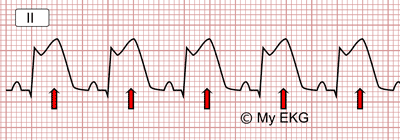


St Segment Analysis


Www Cmaj Ca Content Cmaj 171 9 1042 Full Pdf


Ecg Learning Center An Introduction To Clinical Electrocardiography


Www Rythmolyon Fr Fiche Adris Pdf Name Common 2fecg athletes consensus 17 Pdf


St Changes Heart Squad
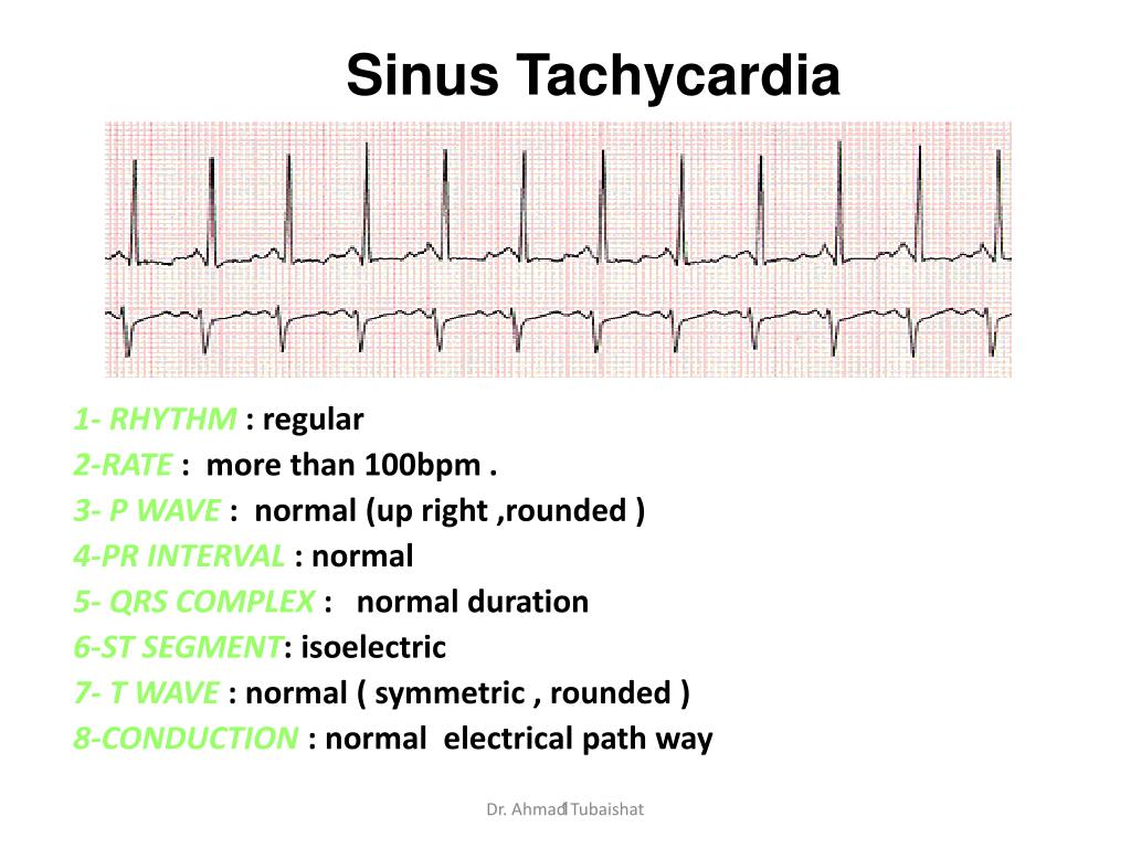


Ppt Sinus Tachycardia Powerpoint Presentation Free Download Id
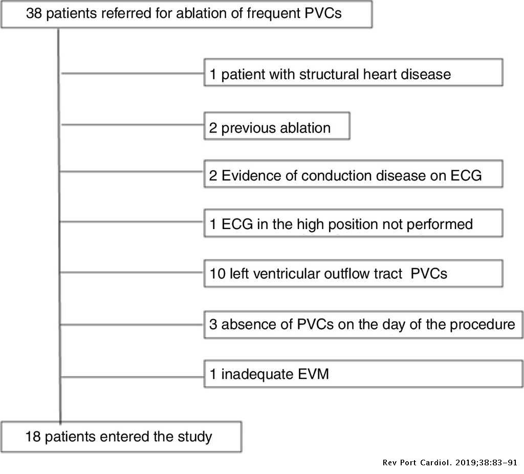


Premature Ventricular Contractions Of The Right Ventricular Outflow Tract Upward Displacement Of The Ecg Unmasks St Elevation In V1 Associated With The Presence Of Low Voltage Areas Revista Portuguesa De Cardiologia


Sinus Tachycardia With No St Segment Depression In The Same Patient Download Scientific Diagram


Ecg Quiz Accelerated Idioventricular Rhythm Arrhythmogenic Right Ventricular Dysplasia Arvd Ashman Phenomenon Asystole Atrial Fibrillation Atrial Flutter Atrial Tachycardia Atrioventricular Block Atrioventricular Dissociation Atrioventricular Nodal


Ecg Fundamentals Of Electrocardiography The Conduction System Is


Acls Rhythm Test 2 A 74 Year Old Woman With Chest Pain Blood Pressure 192 90 And Rates Her Pain 9 10 Pdf Free Download
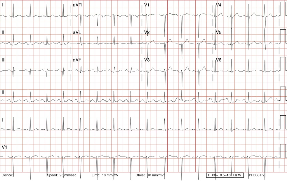


Ii Cardiology Radiology Key


Electrophysiology Of The Heart Ecg Monitoring The Ecg
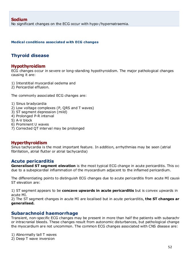


The Normal Ecg And The 12
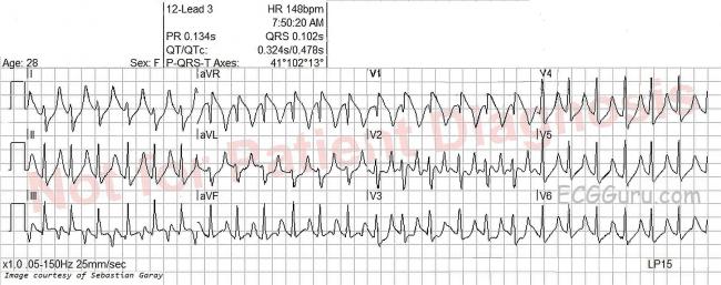


Sinus Tachycardia Ecg Guru Instructor Resources


2


St Segment Elevation And Depressions In Supraventricular Tachycardia Without Coronary Artery Disease
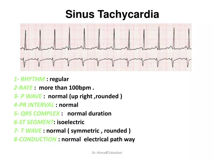


Ppt Sinus Tachycardia Powerpoint Presentation Free Download Id


Sinus Tachycardia With No St Segment Depression In The Same Patient Download Scientific Diagram


Acute Myocardial Infarction Nursingce


Www Ahajournals Org Doi Pdf 10 1161 Jaha 1 Download True


Ekg Medic Rhythms Atrial And Vent Flashcards Quizlet


コメント
コメントを投稿